You are here: Urology Textbook > Urologic surgery > Paramedian laparotomy
Paramedian Laparotomy: Surgical Steps
Urologic Indications for a Paramedian Laparotomy
- A paramedian incision is used for an extraperitoneal approach to the distal and mid-ureter.
- A transperitoneal approach to the abdominal cavity is also possible with a paramedian incision as an alternative to a median laparotomy (without noticeable benefits).
 |
Patient positioning:
Supine position with slight hyperextension of the lumbar spine.
Surgical Technique of a Paramedian Laparotomy
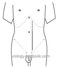
A paramedian skin incision 3–5 cm off the midline and a paramedian incision of the anterior lamina of the rectus sheath is done. Dissect the tendinous intersections from the rectus sheath's anterior lamina until the rectus muscle's medial edge is seen. The rectus muscle is pulled to the lateral by a retractor. For an extraperitoneal approach, a paramedian incision of the fascia transversalis (below arcuate line) and posterior lamina of the rectus sheath (above arcuate line) without incision of the peritoneum is done. Blunt dissection develops the retropubic space, and the peritoneum is dissected to the medial, beginning from caudal to cranial.
Wound closure:
Close the rectus sheath in two layers (anterior and posterior sheath) with continuous running suture (monofilament, elastic, slowly absorbable, suture size USP 0 or 2-0), tissue bites of 5 mm and intersuture spacing of 4--5 mm are applied exclusively to the fascia (Deerenberg et al., 2015).
| Pfannenstiel incision | Index | Gibson incision |
Index: 1–9 A B C D E F G H I J K L M N O P Q R S T U V W X Y Z
References
E. B. Deerenberg et al., “Small bites versus large bites for closure of abdominal midline incisions (STITCH): a double-blind, multicentre, randomised controlled trial.,” Lancet, vol. 386, no. 10000, pp. 1254–1260, 2015, doi: 10.1016/S0140-6736(15)60459-7.
J. A. Smith, S. S. Howards, G. M. Preminger, and R. R. Dmochowski, Hinman’s Atlas of Urologic Surgery Revised Reprint. Elsevier, 2019.
 Deutsche Version: Paramediane Laparotomie
Urology-Textbook.com – Choose the Ad-Free, Professional Resource
This website is designed for physicians and medical professionals. It presents diseases of the genital organs through detailed text and images. Some content may not be suitable for children or sensitive readers. Many illustrations are available exclusively to Steady members. Are you a physician and interested in supporting this project? Join Steady to unlock full access to all images and enjoy an ad-free experience. Try it free for 7 days—no obligation.
New release: The first edition of the Urology Textbook as an e-book—ideal for offline reading and quick reference. With over 1300 pages and hundreds of illustrations, it’s the perfect companion for residents and medical students. After your 7-day trial has ended, you will receive a download link for your exclusive e-book.