You are here: Urology Textbook > Anatomy > Abdominal cavity
Anatomy of the Abdominal Cavity: Muscles
- Anatomy of the abdominal cavity: Muscles
- Anatomy of the abdominal cavity: Nervous system
- Anatomy of the abdominal cavity: Arteries
- Anatomy of the abdominal cavity: Veins and lymphatic system
Muscles of the Abdominal Wall
Ventral and Lateral Abdominal Wall Muscles
Rectus Abdominis Muscle:
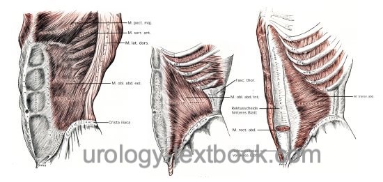
Latin: Musculus rectus abdominis. Long, flat, paired muscle. Origin from the ribs and proc. xiphoideus. Insertion at the pubic bone [fig. ventral abdominal muscles right].
 |
Rectus Sheath
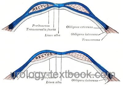
The rectus abdominis muscle is embedded within the aponeurosis of the lateral abdominal muscles, the rectus sheath. The cranial part of the rectus sheath, above the arcuate line, the aponeuroses divide into a anterior and posterior laminae of the rectus sheath. The caudal part of the rectus sheath, below the arcuate line, all parts of the aponeuroses pass in front of the rectus muscle. For the exact anatomy of the rectus sheath, see fig. rectus sheath.
 |
External Oblique Abdominal Muscle:
Latin: M. obliquus externus abdominis, most superficial of the three flat muscles of the lateral and anterior abdominal wall. Origin from the 6th to 12th ribs. Insertion at the rectus sheath and linea alba [fig. ventral abdominal muscles left].
Internal Oblique Abdominal Muscle:
Latin: M. Obliquus internus abdominis. Muscle layer of the anterior and lateral abdominal wall layered in the middle between the external oblique muscle and transverse abdominal muscle. Origin from the inguinal ligament, iliac crest and the lumbodorsal fascia. Insertion: 9th to 12th ribs, rectus sheath and linea alba [fig. ventral abdominal muscles center].
Transverse Abdominal Muscle:
Latin: M. transversus abdominis. Inner muscle layer of the anterior and lateral abdominal wall layered below the internal oblique muscle. Origin from ribs 6-12, fascia thoracolumbalis, and crista iliaca. On the posterior and posterior sheath of the rectus sheath [fig. ventral abdominal muscles right].
Cremaster muscle:
Latin: M. cremaster. Thin layer of skeletal muscle between the external and internal layers of the spermatic fascia, surrounding the testis and spermatic cord. Origin: internal oblique abdominal muscle. The muscle contraction elevates the testes towards the inguinal canal (cremaster reflex).
Dorsal Muscles of the Abdominal Wall
Quadratus Lumborum Muscle:
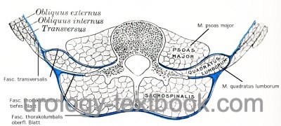
Latin: M. quadratus lumborum. Origin from the 12th rib and transverse processes of the lumbar spine, insertion to the iliac crest [fig. dorsal abdominal wall muscles].
Psoas Major Muscle:
Latin: M. psoas major.The psoas muscle forms together with the spine the dorsal boundary of the abdominal cavity. Origin at the vertebral column. The psoas muscle, together with the iliac muscle, passes through the muscular lacuna to the thigh, and acts as a hip flexor by its attachment to the lesser trochanter of the femur. On the ventral side of the psoas muscle, the tendon of the psoas minor muscle may be present in 30-50% (fig. dorsal abdominal wall muscles].
 |
| Catecholamines | Index | Abdominal anatomy |
Index: 1–9 A B C D E F G H I J K L M N O P Q R S T U V W X Y Z
References
Benninghoff 1993 BENNINGHOFF, A.: Makroskopische Anatomie, Embryologie und Histologie des Menschen.15. Auflage.
München; Wien; Baltimore : Urban und Schwarzenberg, 1993
 Deutsche Version: Anatomie Abdomen
Urology-Textbook.com – Choose the Ad-Free, Professional Resource
This website is designed for physicians and medical professionals. It presents diseases of the genital organs through detailed text and images. Some content may not be suitable for children or sensitive readers. Many illustrations are available exclusively to Steady members. Are you a physician and interested in supporting this project? Join Steady to unlock full access to all images and enjoy an ad-free experience. Try it free for 7 days—no obligation.
New release: The first edition of the Urology Textbook as an e-book—ideal for offline reading and quick reference. With over 1300 pages and hundreds of illustrations, it’s the perfect companion for residents and medical students. After your 7-day trial has ended, you will receive a download link for your exclusive e-book.