You are here: Urology Textbook > Examinations > CT scan
Computed Tomography (CT-Scan) – Technique and Sectional Anatomy
Indications in Urology
Abdominal CT Scan:
CT scan of the abdomen is becoming the preferred radiological imaging technique in urology and is replacing intravenous urography for most indications (Joudi et al., 2006).
Non-Contrast CT:
Computed tomography without contrast medium is the standard imaging technique for patients with renal colic. In addition to the reliable detection of nephrolithiasis and urinary obstruction, non-contrast CT can detect many important differential diagnoses such as cholelithiasis, bowel perforation, aortic aneurysm, ileus, or diverticulitis. Another key advantage is the avoidance of contrast medium.
Contrast-enhanced CT:
A contrast-enhanced CT scan is indicated for the diagnosis of hydronephrosis, hematuria, diagnosis and staging of urogenital tumors, abdominal trauma with suspected renal injury, adrenal tumors, and diseases of the renal vessels.
Further indications for CT in urology:
CT of the chest (pulmonary metastasis, pulmonary embolism), cranial CT (brain metastasis), and CT of the spine or pelvis (to assess the stability of bone metastases).
Examination Technique of CT Scan
Generation of the CT Scan Image:
A thin X-ray beam is generated on one side of the patient and detected on the other after passing through the patient. The X-ray generator and the detector are rotated around the patient for each plane. For each volume element of the body (voxel), a gray value is calculated by complicated algorithms using the associated X-ray attenuations. Early CT devices delivered only sectional images in transverse planes.
Hounsfield scale:
The radiopacity of the voxels is imaged using a gray value calculated with the help of the Hounsfield scale using Hounsfield units (HU): air is by definition -1000 HU, fat varies between -200 to -20 HU, water is by defintion 0 HU, muscle 20–50 HU and cortical bone over 1000 HU. Since the human eye cannot differentiate thousands of gray values, the range of gray values is limited or stretched depending on the clinical question and organ system (windowing technique, gray level mapping).
Image processing:
Modern computer tomographs use multi-line detectors to achieve a higher examination speed. This improves the longitudinal resolution, reduces motion artifacts, and enables several image processing options.
Multiplanar reformation (MPR):
Sagittal and frontal reconstructions can be calculated with a comparable resolution as transverse sectional images [fig. Multiplanar reformation (MPR)]. Curved reconstructions are used to visualize the path of anatomical structures like ureters or blood vessels.
| Do you want to see the illustration? Please support this website with a Steady membership. In return, you will get access to all images and eliminate the advertisements. Please note: some medical illustrations in urology can be disturbing, shocking, or disgusting for non-specialists. Click here for more information. |
Maximum intensity projection (MIP):
The three-dimensional image data set (of maximum intensity of a specific region) is projected onto a two-dimensional plane, thus imitating conventional imaging. Examples: CT angiography with MIP imaging in the frontal plane imitates conventional angiography, or MIP imaging of kidney stones in the frontal plane imitates the X-ray abdomen [fig. Maximum intensity projection].
| Do you want to see the illustration? Please support this website with a Steady membership. In return, you will get access to all images and eliminate the advertisements. Please note: some medical illustrations in urology can be disturbing, shocking, or disgusting for non-specialists. Click here for more information. |
3D Volume rendering (VR):
Complex image processing with three-dimensional imaging of organs, tumors or virtual cast specimens of hollow organs [fig.~\ref{nierenstein_ct_vrt}].
| Do you want to see the illustration? Please support this website with a Steady membership. In return, you will get access to all images and eliminate the advertisements. Please note: some medical illustrations in urology can be disturbing, shocking, or disgusting for non-specialists. Click here for more information. |
Cine mode:
The image processing methods mentioned above can also be displayed in film-like sequences, enabling a rotational view of complex structures.
CT of the Kidneys:
The best diagnostic accuracy is obtained using multiple contrast phases and a high-resolution multidetector CT (MDCT). Unfortunately, this results in a significantly high radiation dose of 20–30 Sv. Depending on the clinical question, some below-mentioned phases may be omitted to spare excess radiation dose.
Non-Contrast Phase:
The non-contrast phase is indicated to detect urinary stones and to measure Hounsfield units of suspicious renal masses or adrenal masses. A reduced radiation dose is often sufficient for a non-contrast CT scan, which yields a minor but sufficient quality in contrast and imaging resolution.
Corticomedullary phase (30–50 s after injection of contrast medium):
Imaging of the arteries and renal cortex with minimal opacification of the renal medulla.
Nephrographic phase (100–160 s after injection):
Imaging of the renal parenchyma and renal vein.
Excretion phase (5–7 min after injection):
Imaging of the collecting system, ureter and bladder (CT urography).
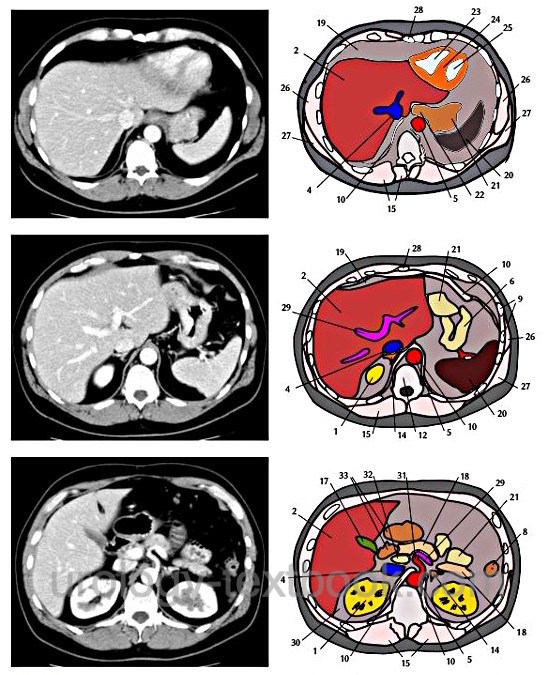
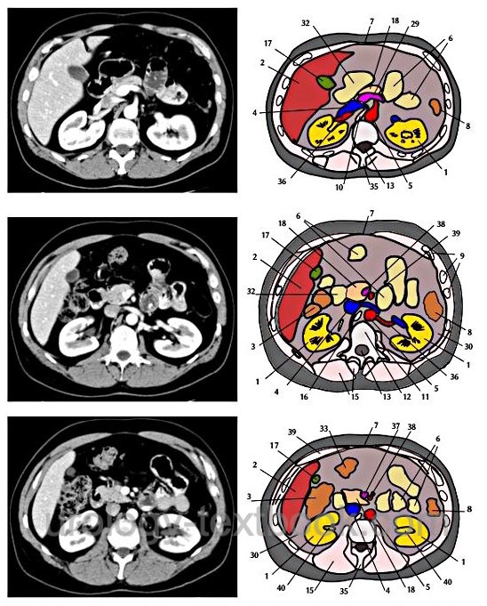
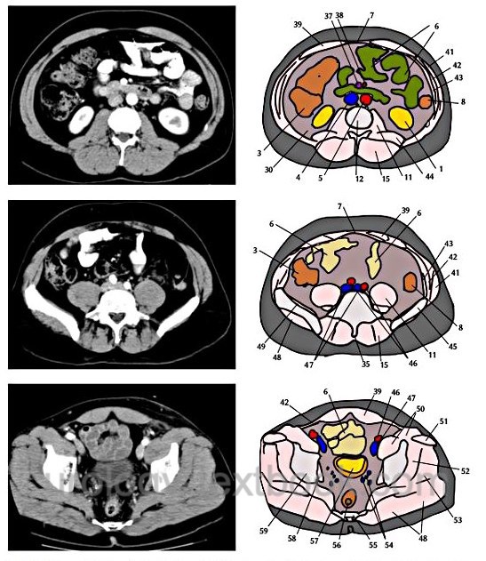
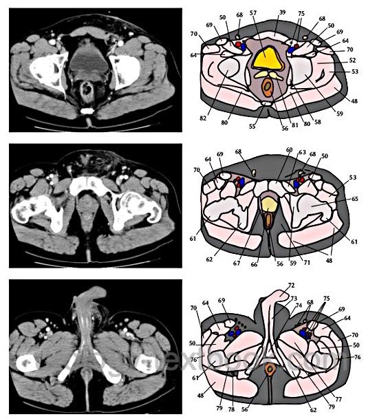
Normal Findings and Sectional Anatomy of the Abdomen and Pelvis CT Scans
Please refer to the following figures for normal sectional anatomy.
 |
 |
 |
 |
| Urologic Surgery | Index | Abbreviations |
Index: 1–9 A B C D E F G H I J K L M N O P Q R S T U V W X Y Z
References
 Deutsche Version: Computer Tomographie (CT): Schnittbild Anatomie des Abdomens
Urology-Textbook.com – Choose the Ad-Free, Professional Resource
This website is designed for physicians and medical professionals. It presents diseases of the genital organs through detailed text and images. Some content may not be suitable for children or sensitive readers. Many illustrations are available exclusively to Steady members. Are you a physician and interested in supporting this project? Join Steady to unlock full access to all images and enjoy an ad-free experience. Try it free for 7 days—no obligation.
New release: The first edition of the Urology Textbook as an e-book—ideal for offline reading and quick reference. With over 1300 pages and hundreds of illustrations, it’s the perfect companion for residents and medical students. After your 7-day trial has ended, you will receive a download link for your exclusive e-book.