You are here: Urology Textbook > Signs and symptoms > Inguinal lymphadenopathy
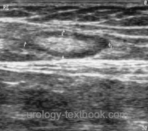
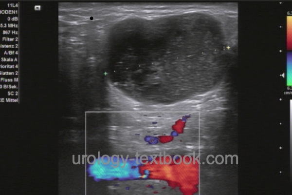
Inguinal Lymphadenopathy: Differential Diagnosis of Groin Lumps
 |
 |
Differential Diagnosis of Inguinal Lymphadenopathy
- Reactive lymph node enlargement
- Genital infections: syphilis, genital herpes, chancroid, lymphogranuloma venereum, granuloma inguinale.
- Malignant tumors: lymphoma, leukemia, penile cancer, melanoma of the penis or lower extremity.
- Systemic infections: mononucleosis, HIV and AIDS, CMV, measles, ...
- Sarcoidosis
| Signs + Symptoms | Index | STD |
Index: 1–9 A B C D E F G H I J K L M N O P Q R S T U V W X Y Z
References
 Deutsche Version: Differentialdiagnose der inguinalen Lymphadenopathie
Urology-Textbook.com – Choose the Ad-Free, Professional Resource
This website is designed for physicians and medical professionals. It presents diseases of the genital organs through detailed text and images. Some content may not be suitable for children or sensitive readers. Many illustrations are available exclusively to Steady members. Are you a physician and interested in supporting this project? Join Steady to unlock full access to all images and enjoy an ad-free experience. Try it free for 7 days—no obligation.