You are here: Urology Textbook > Bladder > Urachal anomalies
Patent Urachus and Urachal Cysts: Diagnosis and Treatment
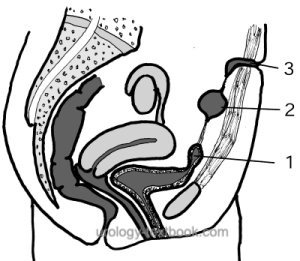
In fetal development, the bladder is connected via the urachus and umbilical cord to the allantois. Usually, the allantois obliterates, leaving the urachus as a fibrous strand at the anterior abdominal wall. Depending on the failure of the regression, various urachal remnants may persist [fig. patent urachus and urachal cysts].
 |
Epidemiology:
In children, the prevalence for sonographic abnormalities is approximately 1:500 (Keceli et al., 2021). The prevalence decreases with age, suggesting a marked spontaneous regression of urachal remnants.
Patent Urachus
A patent urachus leads to postpartum persistent loss of urine via the urachus at the umbilicus. Therapy consists of resection of the urachal fistula. Alternatively, a conservative attempt with urinary bladder catheterization and waiting for spontaneous closure is possible (Lipskar et al., 2010).
Insufficient closure of the urachus:
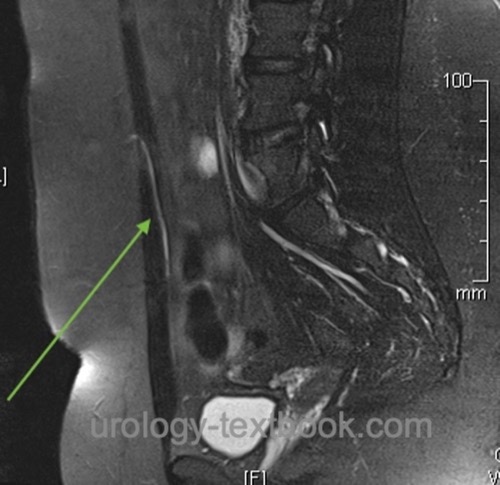
If the urachus obliterates only at the level of the umbilicus, a vesicourachal diverticulum develops near the bladder apex. The vesicourachal diverticulum may lead to urinary tract infections and bladder stones. If the urachus obliterates only at the level of the bladder, an umbilical-urachus sinus develops with an opening at the umbilicus. The umbilical-urachus sinus causes continuous (purulent) draining [fig. mri of an umbilical-urachus sinus].
 |
Urachal cysts
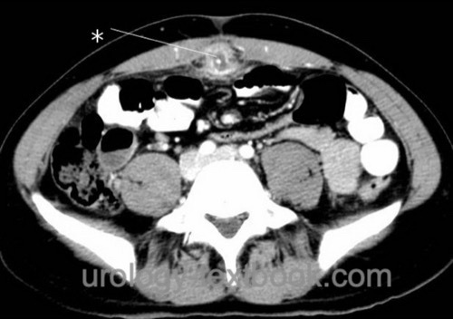
A urachal cyst develops if the urachus obliterates at the level of the bladder and umbilicus but not in between. Urachal cysts may become symptomatic in adults with cyst infection [fig. superinfected urachal cyst] or as a palpable mass. The treatment of symptomatic urachal cysts consists of either percutaneous drainage or surgical (laparoscopic) cyst excision (Lipskar et al., 2010).
 |
Diagnosis of Urachal Remnants:
Ultrasound imaging, VCUG, MRI, cystoscopy.
Treatment
Surgical treatment is necessary only for symptomatic urachal remnants. The risk of developing a urachal carcinoma is considered very low (1:5000); this does not justify the prophylactic resection of visible urachal remnants (Gleason et al., 2015).
| Bladder physiology | Index | Bladder duplication |
Index: 1–9 A B C D E F G H I J K L M N O P Q R S T U V W X Y Z
References
J. M. Gleason et al., “A comprehensive review of pediatric urachal anomalies and predictive analysis for adult urachal adenocarcinoma.,” J Urol, vol. 193, no. 2, pp. 632–636, 2015.
Keç, “Are urachal remnants really rare in children? An observational study.,” Eur J Pediatr, vol. 180, no. 6, pp. 1987–1990, 2021.
Lipskar, A. M.; Glick, R. D.; Rosen, N. G.; Layliev, J.; Hong, A. R.; Dolgin, S. E. & Soffer, S. Z.
Nonoperative management of symptomatic urachal anomalies.
J Pediatr Surg, 2010, 45, 1016-1019
 Deutsche Version: Fehlbildungen des Urachus'