You are here: Urology Textbook > Kidneys > Acute pyelonephritis & Symptoms and diagnosis
Acute Pyelonephritis: Signs, Symptoms and Diagnosis
- Acute pyelonephritis: definition, causes and pathology
- Acute pyelonephritis: symptoms and diagnostic workup
- Acute pyelonephritis: treatment
Guidelines: EAU Guidelines Urological Infections, (German S3 Guidelines UTI).
Signs and Symptoms of Acute Pyelonephritis
- Sudden fever, chills, malaise, and weakness
- Constant flank pain. Children often complain about abdominal pain.
- Flank tenderness
- Frequency, dysuria, (micro)-hematuria
- Possible symptoms: nausea, vomiting, diarrhea, abdominal tenderness, decreased bowel sounds.
- Tachycardia, hypotension, and further symptoms of urosepsis
Complications:
- Renal scarring, especially in children, with the development of chronic pyelonephritis
- Renal abscess
- Urosepsis with septic shock
- Emphysematous pyelonephritis in diabetes mellitus (high mortality)
Diagnosis of Acute Pyelonephritis
The diagnosis is mainly based on the triad of fever, flank pain, and symptoms of bacterial cystitis. Radiological signs are discreet and ambiguous, they are found only in every fourth patient. The clinical value of imaging lies in the detection of complications and for differential diagnosis (Dalla-Palma and Pozzi-Mucelli, 2000) (Kawashima et al., 2000).
Blood Tests:
- Leukocytosis
- Elevated ESR, elevated CRP
- Blood culture: pathogen can be detected in severe disease (high fever, signs of urosepsis)
Urine Sediment:
- Pyuria: cloudy urine, positive dipstick leukocyte esterase test, granulocytes (and white cell casts) in microscopic examination
- Proteinuria
- Microhematuria is typical, sometimes visible hematuria
Urine Culture:
Before antibiotic treatment, a urine culture is always indicated for identification and resistance testing of the responsible pathogen.
Ultrasonography of the Kidneys:
Ultrasonography of the kidneys is indicated for the exclusion of upper urinary tract obstruction. Sonographic signs of pyelonephritis are nonspecific and only valid compared to previous imaging: renal enlargement and hypoechoic parenchyma.
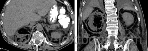
In emphysematous pyelonephritis, the trapped air produces echogenic structures with posterior acoustic shadowing, which are distributed in the parenchyma and perirenal fat. A renal abscess presents as hypoechoic mass, which may contain air. Refer the patient to a CT scan if signs of air or abscess formation are detected.
Computed Tomography:
Contrast-enhanced CT is indicated if renal abscess, nephrolithiasis, emphysematous pyelonephritis or urinary tract obstruction is suspected or if no adequate treatment effect is observed within 2–3 days of proper antibiotic treatment.
CT reliably detects above mentioned complications. The signs of uncomplicated pyelonephritis are subtle: kidney enlargement, wedge-shaped regional limitation of enhancement, delayed nephrogram, perirenal inflammatory infiltrates, and decreased renal function. If retention parameters are elevated, non-contrast spiral CT is an excellent alternative to detect complications.
 |
Intravenous Urography:
Intravenous urography is (was) indicated if urinary obstruction or urinary stones are suspected. Nowadays, intravenous urography is replaced with computed tomography.
Radiological signs of acute PN in urography are discreet and ambiguous: unilateral renal enlargement, delayed enhancement of the affected kidney, and slightly spread renal calyces (by the swollen parenchyma). Ureteropyelitis may be visible by ectasia or by streaks of mucosa due to edema. Destructive stages of (chronic) pyelonephritis may show renal atrophy and papillary destruction or necrosis. In emphysematous pyelonephritis, urography may show trapped gas within the renal fascia. In these cases, the kidney has usually a poor function and urinary obstruction cannot be excluded, CT is recommended. Gas within the collecting system is less dramatic and should not be confused with emphysematous pyelonephritis.
Diagnosis of vesicoureteral reflux:
Recurrent pyelonephritis requires the exclusion or diagnosis of vesicoureteral reflux, particularly in children. Renal ultrasound imaging is done first. In boys older than one year and adults, normal renal ultrasound findings do not warrant further tests. In girls and children under 12 months, MCU and/or DMSA scintigraphy should be performed. It is controversial, if MCU or DMSA kidney scintigraphy is done first or if both examinations are necessary. In adults, the risk for significant VUR after uncomplicated pyelonephritis is low (2%), and MCU or scintigraphy is not recommended.
Differential Diagnosis
- Pancreatitis
- Basal pneumonia
- Pleuritis
- Acute appendicitis
- Acute cholecystitis
- Diverticulitis
- Pelvic inflammatory disease
- Renal and perirenal abscess.
| Pyelonephritis | Index | PN treatment |
Index: 1–9 A B C D E F G H I J K L M N O P Q R S T U V W X Y Z
References
G. Bonkat, R. Bartoletti, F. Bruyère, S. E. Geerlings, F. Wagenlehner, and B. Wullt, “EAU Guideline: Urological Infections.” [Online]. Available: https://uroweb.org/guidelines/urological-infections/
DGU, DEGAM, and PEG, “S3 Leitlinie Epidemiologie, Diagnostik, Therapie, Prävention und Management unkomplizierter, bakterieller, ambulant erworbener Harnwegsinfektionen bei erwachsenen Patienten Aktualisierung 2024.” [Online]. Available: https://register.awmf.org/assets/guidelines/043-044l_S3_Epidemiologie-Diagnostik-Therapie-Praevention-Management-Harnwegsinfektione-Erwachsene-HWI_2024-09.pdf
Dalla-Palma und Pozzi-Mucelli 2000 DALLA-PALMA, L. ; POZZI-MUCELLI, F.: [The imaging of chronic renal infections].In: Radiologe
40 (2000), Nr. 6, S. 537–46
Fihn 2003 FIHN, S. D.:
Clinical practice. Acute uncomplicated urinary tract infection in
women.
In: N Engl J Med
349 (2003), Nr. 3, S. 259–66
Kawashima u.a. 2000 KAWASHIMA, A. ; SANDLER,
C. M. ; GOLDMAN, S. M.:
Imaging in acute renal infection.
In: BJU Int
86 Suppl 1 (2000), S. 70–9
Nickel 2001 NICKEL, J. C.:
The management of acute pyelonephritis in adults.
In: Can J Urol
8 Suppl 1 (2001), S. 29–38
Roberts 1999 ROBERTS, J. A.:
Management of pyelonephritis and upper urinary tract infections.
In: Urol Clin North Am
26 (1999), Nr. 4, S. 753–63
 Deutsche Version: Klinik und Diagnose der Pyelonephritis
Urology-Textbook.com – Choose the Ad-Free, Professional Resource
This website is designed for physicians and medical professionals. It presents diseases of the genital organs through detailed text and images. Some content may not be suitable for children or sensitive readers. Many illustrations are available exclusively to Steady members. Are you a physician and interested in supporting this project? Join Steady to unlock full access to all images and enjoy an ad-free experience. Try it free for 7 days—no obligation.
New release: The first edition of the Urology Textbook as an e-book—ideal for offline reading and quick reference. With over 1300 pages and hundreds of illustrations, it’s the perfect companion for residents and medical students. After your 7-day trial has ended, you will receive a download link for your exclusive e-book.