You are here: Urology Textbook Examinations > Scintigraphy
Renal Scintigraphy: Indications and Normal Findings
Principles of Scintigraphy
Imaging is based on radiopharmaceuticals (tracers) that emit gamma rays. Technetium (99mTc) is the preferably used radioactive marker. The tracers accumulate in particular cells or fluids, depending on their chemical and biological properties, or are excreted via the kidneys. The emitted radiation is detected with a gamma camera and converted into an image. The detection of gamma rays is possible with a scintillation crystal, which generates light flashes when gamma quanta hit the crystal. The light flashes are converted into an electronic signal and imaged according to the intensity as a pixel in different gray values or colors.
 |
Static Renal Scintigraphy
Static renal scintigraphy uses 99mTc-labeled dimercaptosuccinic acid (DMSA). DMSA accumulates in the renal cortex and is scanned 3–6 h after administration. DMSA scintigraphy detects sensitively functioning kidney tissue.
Indications of Static Renal Scintigraphy:
- Imaging of parenchyma scars in vesicoureteral reflux
- Determination of split renal function in chronic kidney disease
- Detection of ectopic kidney tissue
Dynamic renal scintigraphy:
Dynamic (or diuretic) renal scintigraphy uses glomerular filtered 99mTc-DTPA (diethylenetriamine pentaacetate inulin analog) or tubular secreted 99mTc-MAG3 (mercaptoacetyl triglycine) as radiopharmaceuticals. Dynamic scintigraphy determines glomerular filtration rate (GFR), morphological changes, urinary tract obstruction, and urogenital tract injury.
DTPA renal scintigraphy:
DTPA is eliminated via glomerular filtration. DTPA scintigraphy measures glomerular filtration rate (GFR) and renal perfusion. The variable plasma protein binding of DPTA may underestimate GFR.
MAG3 renal scintigraphy:
MAG3 is eliminated to 90% via tubular secretion, 10% by the hepatobiliary system. MAG3 scintigraphy cannot measure GFR, renal function is estimated with renal perfusion (see below). Later images determine the passage of the urine and objectify urinary obstruction. MAG3 has the advantage of a higher activity yield than DTPA and is more suitable than DTPA to measure urinary tract obstruction.
Normal findings of Dynamic Renal Scintigraphy:
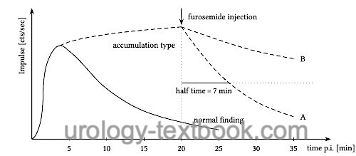
The normal renogram has three phases. The first phase (perfusion phase) shows a rapid increase in activity in the kidney region and correlates with the perfusion of the kidneys. The second phase (secretion or renal phase) shows the peak of activity increase, which is reached after 2–5 minutes. In the third phase (excretion phase), there is a gradual loss of activity in the kidney region due to urinary transport and increased activity in the bladder area. The half-life of activity in the excretion phase is usually below 10 min (Zajic et al., 2004).
If the excretion phase reveals obstruction, urinary excretion is stimulated with a furosemide injection. An enlarged upper urinary tract may mimic urinary obstruction with initial tracer retention. After 20 minutes, at least 50% of the activity should be eliminated after the furosemide injection. Otherwise, the urinary obstruction should be considered detrimental to renal function [fig. findings in dynamic renal scintigraphy].
Captopril renal scintigraphy:
Captopril renal scintigraphy is used to evaluate renal artery stenosis. Renal function declines after injection of captopril if a significant renal artery stenosis is present. Renal scintigraphy is done before and 1 h after captopril 25 mg per os or enalapril 0,04 mg/kgBW IV Any ACE inhibitor medication must be paused before the test.
Bone Scintigraphy
In urology, bone scintigraphy is used to search for bone metastases due to prostate carcinoma, renal cell carcinoma, and bladder cancer or upper tract urothelial carcinoma. The tracers are diphosphonate compounds labeled with 99mTc. Bone scintigraphy is a very sensitive method for the detection of bone metastases. However, suspicious lesions must be confirmed with plain x-ray, CT scan, or MRI since many diseases (old fractures, degeneration, arthritis) may cause false positive results.
Scintigraphy of Pheochromocytoma
Pheochromocytomas or their metastases can be identified with 123iodine or 131iodine labeled meta-iodobenzylguanidine (MIBG) scintigraphy. In addition, paragangliomas or tumors of the APUD system are also visualized.
| MRI | Index | PET scan |
Index: 1–9 A B C D E F G H I J K L M N O P Q R S T U V W X Y Z
References
T. Zajic and E. Moser “[Procedure guidelines for dynamic renal scintigraphy].,” Nuklearmedizin: vol. 43, no. 5, pp. 177–180, 2004.
 Deutsche Version: Technik, Indikationen und Normalbefunde der Nierenszintigraphie
Urology-Textbook.com – Choose the Ad-Free, Professional Resource
This website is designed for physicians and medical professionals. It presents diseases of the genital organs through detailed text and images. Some content may not be suitable for children or sensitive readers. Many illustrations are available exclusively to Steady members. Are you a physician and interested in supporting this project? Join Steady to unlock full access to all images and enjoy an ad-free experience. Try it free for 7 days—no obligation.
New release: The first edition of the Urology Textbook as an e-book—ideal for offline reading and quick reference. With over 1300 pages and hundreds of illustrations, it’s the perfect companion for residents and medical students. After your 7-day trial has ended, you will receive a download link for your exclusive e-book.