You are here: Urology Textbook > Ureters > Retrocaval ureter
Diagnosis and Treatment of a Retrocaval Ureter
Definition of Retrocaval Ureter
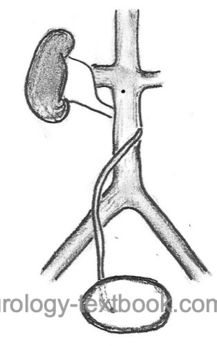
A retrocaval ureter, also called a circumcaval ureter, is an abnormal development of the vena cava and ureter. Due to the persistence of the right subcardinal vein in the lumbar area, the vena cava develops ventrally of the right ureter (Zhang et al., 1990).
 |
Epidemiology
1:1500
Etiology of Retrocaval Ureter
The ureter lies between the following fetal veins: dorsally, the supracardinal vein, and posterior cardinal vein; ventrally, the subcardinal vein. Usually, the portion caudal of the renal vein of the inferior vena cava develops from the right supracardinal vein, and the ureter lies anterior to the inferior vena cava. If the subcardinal vein persists, the inferior vena cava develops ventral of the ureter and may cause hydronephrosis by compression between vena cava and spine.
Signs and Symptoms
- Right-sided flank pain
- Nephrolithiasis
- Pyelonephritis
- Loss of right-sided kidney function is possible.
Diagnostic Workup
Intravenous urography:
The right mid-ureter suddenly turns to the medial; there may also be a change in diameter. Caudally from the ureteral kinking, there is often no contrast in the ureter. Thus, the ureter looks like a "J". Signs of hydronephrosis or nephrolithiasis may be present.
Retrograde pyelography:
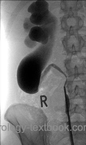
The spiral (S-shaped) curve of the ureter around the vena cava is visible [fig. retrocaval ureter].
Computed tomography or MRI:
The abnormal position of the ureter to the inferior vena cava is easily identified.
 |
Treatment of Retrocaval Ureter
Surgical treatment is necessary for complications, such as intermittent flank pain, kidney stones, recurrent infections, or significant hydronephrosis with loss of kidney function. Surgery includes the division of the ureter after complete mobilization, spatulation of the ureteral ends, and reanastomosis ventral of the vena cava (ureteroureterostomy). The technical difficulty of the operation is the necessity of extensive dissection of adhesions between ureter and vena cava with the corresponding risk of injury and bleeding. If the fibrotic adhesions between the ureter and vena cava are severe, the atretic ureter segment can be left behind the vena cava. Open surgery requires a significant retroperitoneal or transperitoneal approach to the vena cava due to the central position within the abdominal cavity. Laparoscopy can reduce the morbidity of the surgical procedure, see the following figures:
| Do you want to see the illustration? Please support this website with a Steady membership. In return, you will get access to all images and eliminate the advertisements. Please note: some medical illustrations in urology can be disturbing, shocking, or disgusting for non-specialists. Click here for more information. |
| Do you want to see the illustration? Please support this website with a Steady membership. In return, you will get access to all images and eliminate the advertisements. Please note: some medical illustrations in urology can be disturbing, shocking, or disgusting for non-specialists. Click here for more information. |
| Do you want to see the illustration? Please support this website with a Steady membership. In return, you will get access to all images and eliminate the advertisements. Please note: some medical illustrations in urology can be disturbing, shocking, or disgusting for non-specialists. Click here for more information. |
| Do you want to see the illustration? Please support this website with a Steady membership. In return, you will get access to all images and eliminate the advertisements. Please note: some medical illustrations in urology can be disturbing, shocking, or disgusting for non-specialists. Click here for more information. |
| Megaureter | Index | Retroiliac ureter |
Index: 1–9 A B C D E F G H I J K L M N O P Q R S T U V W X Y Z
References
Zhang, X. D.; Hou, S. K.; Zhu, J. H.; Wang, X. F.;
Meng, G. D. & Qu, X. K.
Diagnosis and treatment of retrocaval
ureter.
Eur Urol, 1990, 18, 207-210
 Deutsche Version: Retrokavaler Ureter