You are here: Urology Textbook > Examinations > Retrograde pyelography
Retrograde Pyelography
Abbreviations: RUP (retrograde ureteropyelography), RPG (retrograde pyelography).
Indications
Retrograde pyelography is rarely performed as a primary diagnostic tool; it is an alternative to intravenous urography or abdominal CT scan if contraindications for iodinated contrast medium exist. In principle, it is indicated for nephrolithiasis, hematuria, urinary obstruction, injury or tumors of the upper urinary tract. It is often done for orientation before ureterorenoscopy or ureteral splint insertion or replacement.
Examination Technique
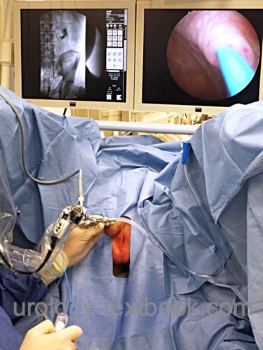
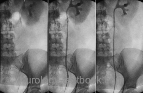
After cystoscopy, a ureteral catheter (5–6 Ch) is inserted into the ostium [fig. RPG] and a baseline KUB without contrast is done [fig. RPG before DJ]. Collimation should be adapted to the examined side (half-sided KUB). The contrast medium is injected slowly using fluoroscopy to judge the injection volume. If the contrast medium does not reach the upper urinary tract in sufficient concentration, the ureteral catheter is advanced into the renal pelvis. After aspiration of urine, contrast medium is injected slowly under fluoroscopy control. The examination is evaluated in real-time with the help of fluoroscopy, significant still images are saved for documentation.
 |
 |
Normal Findings
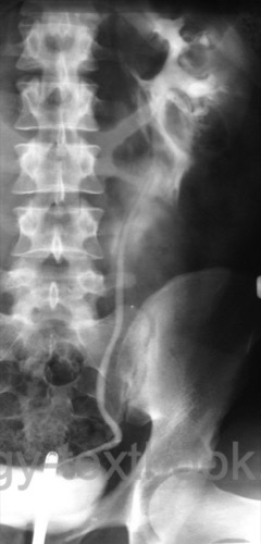
See the section abdominal X-ray for normal findings of the initial KUB (plain film). The retrograde contrast medium injection reveals a slim upper urinary tract; for the normal anatomy, see the previous section intravenous urography, and fig. radiological anatomy of the renal calyces. Sometimes air bubbles mimic filling defects suspicious for a stone or tumor. After removing the ureteral catheter, the contrast medium flows into the bladder.
 |
Complications
Injury of the ureter:
A kinked ureter, ureteral stricture, tumor, or ureteral stone can prevent advancement of the ureteral catheter. Beware of ureteral perforation and advancement of the catheter into the ureteral wall or retroperitoneum. If resistance is encountered, further progress is possible using a hydrophilic-coated guidewire and fluoroscopy.
Forniceal rupture:
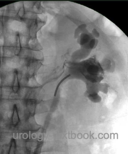
Too rapid injection or too much contrast medium can lead to pyelolymphatic reflux or extravasation of the contrast agent in the area of the fornices. Aspiring urine from the renal pelvis prior to administration of the contrast medium and slow injection under fluoroscopy reduces the probability of a forniceal rupture.
 |
 |
Urosepsis:
Retrograde pyelography can induce fulminant urosepsis in patients with infected hydronephrosis. The aspiration of urine from the renal pelvis prior to application of the contrast medium allows a urine culture and reduces the risk of bacteriemia. contrast medium should be applied as sparingly as possible, an orienting imaging of the renal pelvis is sufficient for the correct positioning of a ureteral splint. The diagnostic workup of the etiology of the obstruction must be postponed to a later date.
Adverse reactions to contrast medium:
The retrograde administration of iodine-containing contrast medium is possible in patients with chronic kidney disease, iodine intolerance or contrast medium allergy. Allergic reactions to the contrast agent are unusual. The risk factor is the injection under pressure with extravasation (Blackwell et al., 2017) (Weese et al., 1993). Comparable to intravenous urography, an emergency kit should be ready to treat anaphylaxis.
| Intravenous urography | Index | VCUG |
Index: 1–9 A B C D E F G H I J K L M N O P Q R S T U V W X Y Z
References
Blackwell, R. H.; Kirshenbaum, E. J.; Zapf, M.
A. C.; Kothari, A. N.; Kuo, P. C.; Flanigan, R. C. & Gupta, G. N.
Incidence
of Adverse Contrast Reaction Following Nonintravenous Urinary Tract
Imaging.
European urology focus, 2017, 3, 89-93.
 Deutsche Version: Retrograde Pyelographie
Urology-Textbook.com – Choose the Ad-Free, Professional Resource
This website is designed for physicians and medical professionals. It presents diseases of the genital organs through detailed text and images. Some content may not be suitable for children or sensitive readers. Many illustrations are available exclusively to Steady members. Are you a physician and interested in supporting this project? Join Steady to unlock full access to all images and enjoy an ad-free experience. Try it free for 7 days—no obligation.
New release: The first edition of the Urology Textbook as an e-book—ideal for offline reading and quick reference. With over 1300 pages and hundreds of illustrations, it’s the perfect companion for residents and medical students. After your 7-day trial has ended, you will receive a download link for your exclusive e-book.