You are here: Urology Textbook > Examinations > Retrograde urethrography
Retrograde Urethrography: Examination Technique and Normal Findings
Indications for Retrograde Urethrography
Retrograde urethrography (RUG) is a X-ray examination of the urethra for evaluation in cases of trauma, diverticula, urethral strictures, urethral fistulas or urethral valves.
Examination Technique for Male Patients:
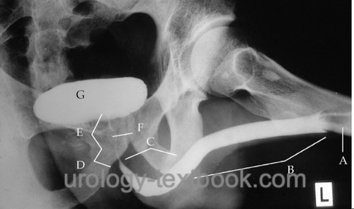
The patient is positioned with an abducted left thigh and raised right pelvis (about 40 degrees) with the examiner standing on the left side of the patient. A penile clamp with injection cannula or a 12 CH catheter is inserted into the urethra, the catheter balloon is inflated with miminal volume in the fossa navicularis [fig. normal retrograde urethrography]. The urethra is straightend up with gentle tension. contrast medium is slowly injected under fluoroscopy. The help of the patient is sometimes necessary to let the contrast medium pass into the bladder. Inspiration, relaxation or Jendrassik handle) help to reduce the resistance of the sphincter.
 |
Normal findings in Men:
The penile and bulbar urethra shows no narrowing. The bulbar urethra ends with a sharp angel into the membranous urethra. The membranous urethra presents with a thin filiforme contrast due to the external sphincter. The prostatic urethra is better contrasted and contrast medium enters the bladder. The seminal colliculus may cause a filling defect of the prostatic urethra [fig. normal retrograde urethrography].
Examination Technique for Female Patients:
The inner and outer meatus of the urethra is sealed with the help of a special double-balloon catheter. The contrast medium is injected slowly under supine or lateral decubitus position.
| VCUG | Index | Retrograde pyelography |
Index: 1–9 A B C D E F G H I J K L M N O P Q R S T U V W X Y Z
References
 Deutsche Version: Retrograde Urethrographie: Röntgen der Harnröhre