You are here: Urology Textbook > Urologic examinations > Imaging > Testicular ultrasound
Testicular Ultrasound Imaging and Doppler Sonography
Testicular and scrotal ultrasound imaging is indicated in patients with testicular pain (acute scrotum), trauma, scrotal masses, male infertility, hypogonadism, gynecomastia and after scrotal surgery. The Doppler ultrasound is additionally performed in patients with an acute scrotum (differential diagnosis of testicular torsion), varicocele and for unclear tumors to detect internal perfusion.
Examination Technique
A high-resolution linear probe of 7.5–10 MHz is used for imaging of the scrotum. In the supine position, the testicles are fixed with a towel or by the hands of the patient. The testicles and scrotal content are systematically examined in longitudinal and transversal sections.
Normal Findings
Testicular size:
In adults: length 4–5 cm, thickness 2–3 cm, volume 12–30 ml. The volume is calculated using the following formula:
Testicular volume = Length × Width × Height × 0,5
Echo architecture:
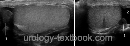
The testicle shows a homogeneous echo pattern, the tunica albuginea is thin and echogenic. The radial arrangement of the lobules can be identified. The epididymis is clearly distinguishable from the testes fig. normal testicular ultrasound.
 |
Doppler Ultrasonography of the Testis
Technical settings:
For a reliable identification of the testicular vessels, a high quality linear probe with a frequency of 10 MHz or above should be used. The settings are set expecting a low flow rate and low Nyquist limit, a wall filter should be turned off.
Normal findings:
It is crucial to perform the Doppler ultrasound examination with unequivocal intratesticular vessels. Capsular vessels may be misidentified as vessels of the scrotal wall. With a correct technical setting, a Doppler signal is always detectable from the testicular parenchyma. Normally, the RI in the testes (resistive index) is below 0.7 due to the relatively high end-diastolic blood flow. Higher RI values (decreasing diastolic blood flow) are signs of partial testicular torsion.
| Penis ultrasound | Index | TRUS |
Index: 1–9 A B C D E F G H I J K L M N O P Q R S T U V W X Y Z
References
Singer u.a. 2006 SINGER, Eric A. ; GOLIJANIN, Dragan J. ; DAVIS, Robert S. ; DOGRA, Vikram: What’s new in urologic ultrasound?In: Urol Clin North Am
33 (2006), Aug, Nr. 3, S. 279–286
 Deutsche Version: Sonographie der Hoden