You are here: Urology Textbook > Urologic examinations > Imaging > ureter ultrasound
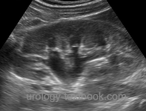
Ureter Ultrasound and Sonography Classification of Hydronephrosis
The indications for ultrasonography of the ureters are comparable to the indications for renal ultrasound (see section renal ultrasound).
 |
Examination Technique
The transducer used for ureter ultrasonography is a sector or curved array probe with 3.5–5 MHz. The examination of the ureters is done in supine position and with inspiration of the patient. In case of unfavorable conditions, a lateral decubitus position is helpful, whereby the side to be examined is raised by 30\textdegree. The kidney is used as an acoustic window, the renal pelvis and the ureteral outlet can be visualized regularly. Intestinal air often prevents the examination of the proximal ureter, the mid portion cannot be examined with ultrasound. The distal part of the ureter is examined in supine position and with full bladder, the bladder is used as an acoustic window.
Hydronephrosis:
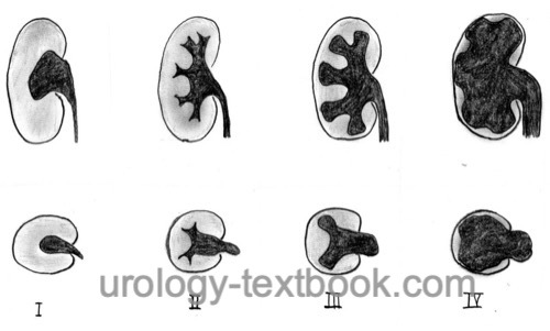
The distinction between hydronephrosis or significant urinary tract obstruction is not possible with sonography. The dilatation of the upper urinary tract is classified according to morphological criteria [fig. classification of hydronephrosis]. Consequently, hydronephrosis should be described in the ultrasonography report without judging the significance of the obstruction (Beetz u.a., 2001).
 |
| Renal ultrasound | Index | Bladder ultrasound |
Index: 1–9 A B C D E F G H I J K L M N O P Q R S T U V W X Y Z
References
Singer u.a. 2006 SINGER, Eric A. ; GOLIJANIN, Dragan J. ; DAVIS, Robert S. ; DOGRA, Vikram: What’s new in urologic ultrasound?In: Urol Clin North Am
33 (2006), Aug, Nr. 3, S. 279–286
 Deutsche Version: Sonographie der Harnleiter
Urology-Textbook.com – Choose the Ad-Free, Professional Resource
This website is designed for physicians and medical professionals. It presents diseases of the genital organs through detailed text and images. Some content may not be suitable for children or sensitive readers. Many illustrations are available exclusively to Steady members. Are you a physician and interested in supporting this project? Join Steady to unlock full access to all images and enjoy an ad-free experience. Try it free for 7 days—no obligation.
New release: The first edition of the Urology Textbook as an e-book—ideal for offline reading and quick reference. With over 1300 pages and hundreds of illustrations, it’s the perfect companion for residents and medical students. After your 7-day trial has ended, you will receive a download link for your exclusive e-book.